- Home
- About Us
- Treatments
- Services
- Doctors
- Patients
- Careers
- Contact Us


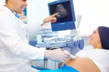
Ultrasound involves the use of high-frequency sound waves to create images of organs and systems within the body. An ultrasound machine creates images that allows various organs in the body to be examined. The machine sends out high-frequency sound waves, which reflect off body structures. The transducer has receptors which receive these signals and convert them into digital signals and form image of the body organ on the computer screen. Ultrasound is the gold standard for looking at the health of foetus in pregnant women. It allows the gynaecologists to detect any congenital abnormalities in the foetus and take corrective action
Ultrasound is extremely useful in diagnosing diseases of the organs in the abdomen, blood vessels and soft tissues. The best thing about ultrasound exam is that it is completely harmless and involves no radiation unlike other diagnostic modalities like X-Ray, CT Scan, Mammography etc.
At Sethi Hospital we have the most advanced Ultrasound and Colour doppler machine from Toshiba Japan. The machine is equipped with Transvaginal Probe. Transvaginal ultrasound usually produces better and clearer images of the female pelvic organs, because the ultrasound probe lies closer to these structures.

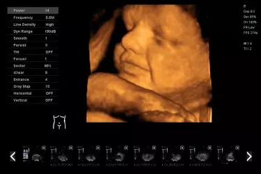
3D/4D ultrasound can obtain views of the pelvis that are not seen on the conventional 2D ultrasound, especially views of the uterus. 2D ultrasound can obtain longitudinal and transverse views of the uterus. 3D ultrasound adds in the coronal view (or C-plane) of the uterus, enabling us to get an image of the uterus that is “front on”.
3D/4D ultrasound may help in the diagnosis and assessment of pelvic conditions, including:
 Congenital uterine abnormalities. Some women are born with changes in the shape of the uterus (for example, bicornuate uterus). Minor changes in the shape of the uterus are not uncommon.
 The location of endometrial polyps.
 The location of submucous fibroids and assessment of any intramural component.
 The location of an intrauterine contraceptive device (IUCD), especially if the IUCD is abnormally located (for example, penetrating into the wall of the uterus).
 Investigation of recurrent miscarriages and preterm deliveries.
The cost of Ultrasound depends upon the type. It ranges from INR 1000 to INR 3000 depending upon the scan type.

Pregnancy Ultrasound - In order to measure the growth of the foetus, ultrasound is the only diagnostic modality. During the nine months of the pregancy atleast 3 ultrasound exams are done, one in every trimester. In the first ultrasound of the pregnancy the viability of the foetus is determined alongwith foetal heart rate and the gestational age. This ultrasound also gives the expected date of delivery.
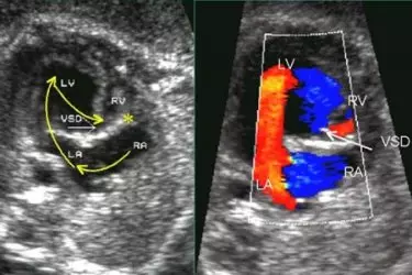
During the second trimester around 14-15 weeks of pregnancy a very detailed and indepth ultrasound is done called Level II Scan which gives complete information about the congenital abnormalities in the foetus.
 Testicle ultrasound
 Thyroid ultrasound
 Transvaginal ultrasound
 Vascular ultrasound

Color Doppler ultrasound is an imaging technique which is used to provide visualization of the blood flow in the arteries and the veins. With the advent of Medical Technology, CT and MRI angiography has emerged as the preferred diagnostic modality for study of blood vessels. In angiography iodine based contrast agent is used which is harmful for kidneys. In comparison a colour doppler study is perfectly safe even for pregnant women as there is no radiation involved and safe for patients of kidney disease as there is no iodine involved.
Colour Doppler is of great use in pregnancy for seeing the position of the umbilical cord, for studying the valves and coronary arteries through 2-D Echocardiography, Renal artery by doing Renal Colour Doppler, Arteries and Veins of the hands and legs to see any blockages in them. Carotid artery for seeing the blockages in the blood going to the brain. The colour doppler study is not only harmless to the body but also a cost effective way of diagnosing the diseases of the blood vessels.
Colour Doppler Studies
![]() Obstetrical Colour Doppler
Obstetrical Colour Doppler
![]() Arterial Doppler
Arterial Doppler
![]() Venous Doppler
Venous Doppler
![]() Penile Doppler
Penile Doppler
![]() Renal Artery Doppler
Renal Artery Doppler
![]() Carotid Doppler
Carotid Doppler
![]() Abdominal Doppler
Abdominal Doppler
![]() 2D Echocardiography
2D Echocardiography
![]() Dobutamine Stress Echo
Dobutamine Stress Echo

At our centres we routinely perform the following tests on the colour doppler or ultrasound machine:
 Find out the blood clots and blocked blood vessels in almost any part of the body
 Evaluate leg pain that may be caused by a condition caused by atherosclerosis of the lower extremities
 Evaluate blood flow after a stroke
 Evaluation of a stroke can be done through a technique called transcranial Doppler (TCD) ultrasound
 Evaluate abnormal veins a variety of ultrasound scans for various organs.
Who Should Get a Doppler Done
 In case your doctor suspects narroawing of carotid arteries then he might ask for a carotid doppler.
 In case doctors suspects of blockage in limbs then he may order a vascular colour doppler.
 In case doctors suspects of blood clots in legs then he may order a venous colour doppler.
 Patients with erectile dysfunction may need a penile doppler to evaluate need for penile implant.
 A gynaecologist would order a pregnancy doppler to see the placental blood flow.
The Cost of Colour Doppler scan varies with the kind of doppler. Cost would range between INR 2500 to 7000 depending upon the study.

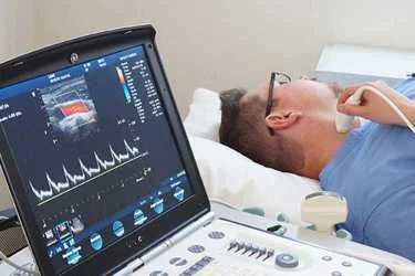
 Carotid Color Doppler - test is used to detect narrowing of the arteries in the neck (the carotid arteries) that supply blood to the brain. The test is known is CIMT - Carotid Intima Mean Thickness. This test is extremely useful in early detection and prevention of future stroke.
 Vascular Color Doppler - Several Medical conditions causes the arteries and veins to become narrow. This reduces the blood supply to different body parts. Arterial and Venous Colour Doppler studies are done mostly for the upper and lower limbs to determine the blockages in the vessels and determine the extent of narrowing of these vessels
 Venous Color Doppler at Siddh Diagnostic Centre in Gurgaon - venous ultrasound exam is to search for blood clots, especially in the veins of the leg. This condition is often referred to as deep vein thrombosis or DVT.
 Transcranial Doppler for children - A transcranial Doppler (TCD) ultrasound evaluates both the direction and velocity of the blood flow in the major cerebral arteries of the brain.
 Penile Colour Doppler - Patients who suffer from erectile dysfunction or impotency can benefit from this study where the blood flow in the penis is studied using Colour Doppler.

Sethi Hospital was setup in the year 1996 by the doctor couple of Dr. Ashok Sethi and Dr Pushpa Sethi in the city of Gurgaon. Initially the hospital offered services in the fields of Orthopaedics and Gynaecology but soon developed into a multi-specialty hospital offering complete range of secondary and tertiary level treatments. In the last 24 years, the hospital has treated more than 12 lac patients throught outdoor and indoor facilities. Sethi Hospital has won the trust of the patients on the basis of ethical and cost effective treatment. Middle class patients who have to self finance their medical treatment, find our pricing quite affordable for the quality of treatment given.

The state of art cutting edge medical technology deployed at Sethi hospital makes us a complete hospital for your medical treatment. Most of the patients who come for any kind of treatment normally require the facilities of ultrasound, colour doppler, echocardiography and digital x-ray within the hospital. Sethi hospital has all these facilities inhouse which means that the patient does not have to run from one place to another for getting the complete treatment. Inhouse pharmacy and pathology lab ensures that the attendants of the patients getting admitted at the hospital can have the peace of mind and do not have to run helter skelter.

The team of doctors at Sethi hospital are some of the most experiencd medical professionals in the city of Gurgaon. Almost all our doctors have more than 15-20 years of experience in their respective fields. The doctors at Sethi Hospital are always trying to improve the medical and surgical outcomes and take active part in continous medical education programmes. Our team of doctors are assisted by highly trained paramedics in the diagnostic field. Our Nursing Team efficiently carries out the doctor's instructions and provide compassionate care which speeds up the healing process. The Physiotherapy Team makes sure that your recovery is quick and complete.
Dr A.K.Sethi, Orthopaedic Surgeon at Sethi Hospital is talking about the surgical procedure for replacement of Knee Joints which are worn due to Osteoarthritis. Watch the Video.
Dr Pushpa Sethi, Gynaecologist is talking about the various treatment options available for Polycystic Ovaries - Lifestyle modification, Diet, Medicines and Surgical.